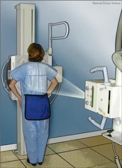
X-rays is a diagnostic medical device which uses X-rays that is an X-ray beam to detect disease. This diagnostic method is still sacrosanct in the initial diagnosis of locomotor system, the diagnosis of pulmonary disease and heart failure, kidney disease and urinary system and gives good results in the study of diseases of the esophagus, stomach and intestines. X-ray machines of new generation with digital camera technology and successfully used in the diagnosis of vascular diseases - angiography.
Each type of radiation carries with it a certain amount of danger. To the danger of X-ray radiation remedy, it is reduced to a minimum, protective measures are implemented during shooting. They include:
· Protection of the environment of X-ray radiation.
Lung X-Ray
Lung X-ray is one of the oldest X-ray method. Viewing is done easily and quickly. The patient is naked to the waist and leaned on board with the film. At the command X-ray technicians patient took a deep breath and hold your breath, after which he made the recording. The procedure takes a total of 5 minutes. X-ray of the lungs was performed at suspected bronchitis, pneumonia, lung tumors, pleural effusions, atelectasis, pneumothorax, pulmonary injury. The detection of these diseases rentgengrafija today represents an unrivaled method.
X-ray examination of the heart (AP radiographs)
This test is performed in the same manner as the examination of the lungs. Rentgengrafijom heart is diagnosed size of cardiac chambers, aneurysms of the thoracic aorta, pleural effusions.
AP radiographs with I and II polukosim
This test is performed as well as X-ray of the heart and lungs, supplemented by two additional footage. On this occasion, the patient should drink a sip of contrast medium-barium and during filming to keep up. Recordings were performed in three positions and instructions on individual positions the patient gives an X-ray technician.
X-rays of bones and joints
All shots locomotor system, filmed part of the body should be released from clothing and jewelry possible. This review is the diagnosis of the following diseases:
· Fracture (fracture) the bone;
· Bone tumors;
· Degenerative diseases;
· Rheumatic diseases of the joints and bones;
· Osteomyelitis (infection of the bone);
· Periostitis (periostitis);
· Bone cysts;
· Congenital anomalies (deformities) locomotor system;
· Determine bone age of the child;
· Paranasal sinuses - sinuses (sinusitis, polyps, etc.).
X-ray of the urinary tract
This review is obtained insight into the appearance (but not function), the size, position and shape of the kidney as well as the possible existence of calculi in the kidneys, ureters or bladder. Viewing is done lying down. This review is generally performed in suspected existence of stones in the kidney or in the ureters or bladder as well as in suspected tumors. On X-rays are analyzed: size, layout and format of the kidneys; the existence of shadows that indicate calculus and look paravertebralene musculature. The whole process only takes 5 minutes.
X-ray of the abdomen
This review is diagnosed ileus (bowel combination) or a possible perforation of the abdominal organs (stomach, intestines). The indications for this review acute abdomen. Viewing is done standing up. This view belongs to a group of emergency medical examination and is usually performed at admission diagnosis 'acute abdomen'.
X-ray of the stomach and esophagus
X-ray examination of the stomach is a method for detecting diseases of esophagus, stomach and duodenum. Viewing is done on an empty stomach. The patient must not eat or drink. During recording, the patient must remove the upper part of their clothing. The patient drinks a contrast agent called barium and then begins review that is recording. Barium has a taste of lemon, a dense content and not a big deal to drink. This review is not painful. During the recording-exposure, it is important that the patient is not breathing and does not move. The examination takes up to 10 minutes. This review does not work with brijumovim milk if suspected bleeding ulcer. In this case, in contrast to the use gastrografin
Barium enema and irigoskopija - X-rays of the colon
X-rays of the colon is a reliable method for detecting diseases of the colon. Barium enema is diagnosed diverticles colon, mucous membrane inflammation of the colon, brain tumors and other diseases. This review is not painful, but it's a little uncomfortable for the patient because the anus must put one thin tube is inserted through which a contrast agent called barium. The patient for this examination must be prepared the previous day (take cleaning).
<Cleaning> evacuation of feces that is performed in the following manner:
· The day before the examination is taken as a light breakfast and a light lunch;
· 16:00 to take some of laksantnih preparations (castor oil or 4 Dulcolax tablets and the like);
· After the examination does not need to eat and to drink water or tea optionally but not too much;
Examination is performed as follows:
· Patient undress completely naked and lie on an examination table (while not recording is covered by a sheet);
· X-ray technician put a small intestine to the anus of the patient and are injected into the anus of barium through the device called pneumokolon. During examination, the patient needs to squeeze the muscles of the anus that would not let barium to get out. The examination takes about 30 minutes;
· During the recording - Exposure patient is not breathing, not moving;
· After this procedure the patient goes to the toilet to empty and record again ...
Intravenous urography
Intravenous urography is a highly reliable method of establishing the function and kidney function, see their appearance as well as the state of renal excretory canal, ureters and bladder. This review provides valuable information in the study arteriske hypertension. In combination with ultrasound represent more than 90% can be relied upon a method for detecting disorders of the kidneys and the bladder. If there is an allergy, an overview is done in the following way:
· The patient lies down on the X-ray table and gets into a vein two vials of contrast medium (60 ml);
· Across the abdomen of the patient to tighten up one broad canvas that engages the patient to the plate x-ray table, and whose task is to prevent the rapid swelling of the contrast agent from the kidneys;
· Thereafter performs recording. I view the last 40 minutes;
· During exposure (recording) the patient can not breathe on it always warns the X-ray technician.